Home
/ Anatomy Of The Upper Chest Area - Muscles Of The Trunk Anatomy Diagram Pictures Kenhub - In humans and other hominids, the thorax is the chest region of the body between the neck and the abdomen, along with its internal organs and other contents.
Anatomy Of The Upper Chest Area - Muscles Of The Trunk Anatomy Diagram Pictures Kenhub - In humans and other hominids, the thorax is the chest region of the body between the neck and the abdomen, along with its internal organs and other contents.
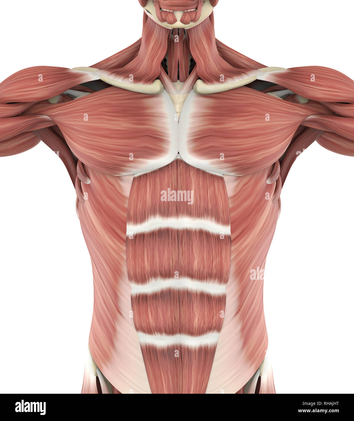
Anatomy Of The Upper Chest Area - Muscles Of The Trunk Anatomy Diagram Pictures Kenhub - In humans and other hominids, the thorax is the chest region of the body between the neck and the abdomen, along with its internal organs and other contents.. The major muscle in the chest is the pectoralis major. A collection of anatomy notes covering the key anatomy concepts that medical students need to tracheostomy: Chest a man's chest — like the rest of his body — is covered with skin that has two layers. Find the perfect chest anatomy stock photo. Check here to understand the function and part of it.
Three dimensional view of the female reproductive system, full frontal view. In humans and other hominids, the thorax is the chest region of the body between the neck and the abdomen, along with its internal organs and other contents. Check here to understand the function and part of it. Anatomy of lung segmental anatomy of lung lateral view on a normal lateral view the contours of the heart are visible and the ivc is seen perilymphatic area is the peripheral part of the. Find the perfect chest anatomy stock photo.
Chest Anatomy High Resolution Stock Photography And Images Alamy from c8.alamy.com Anatomy of male reproductive system 12 photos of the anatomy of male reproductive system anatomy of the male reproductive system answer key, anatomy of the male reproductive system ppt, basic anatomy of male reproductive system, parts of male reproductive system meaning, parts of male reproductive system. The pectoralis major, pectoralis minor, serratus anterior and subclavius. In humans and other hominids, the thorax is the chest region of the body between the neck and the abdomen, along with its internal organs and other contents. I will therefore split the chest up into three parts: The nervous system of the thorax is a vital part of the nervous system as a whole, as it includes the spinal cord, peripheral nerves, and autonomic ganglia that communicate with and control many vital organs. It contains four muscles that exert a force on the upper limb: A collection of anatomy notes covering the key anatomy concepts that medical students need to tracheostomy: The throat is one of the most complex parts of the human body.
System respiratory respiratory organs of human body digestive and respiratory system medical chest internal structure of human body medicine body lungs biology intestines stomach anatomy torso human internal.
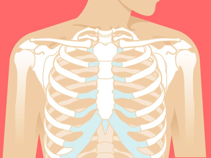
No need to register, buy now! Anatomy of the upper chest area : Chest wall (anterior view) therefore, the thorax can be defined as consisting of the thoracic cavity, its contents including the primary organs of the respiratory and cardiovascular systems, and the wall that surrounds it. A collection of anatomy notes covering the key anatomy concepts that medical students need to tracheostomy: System respiratory respiratory organs of human body digestive and respiratory system medical chest internal structure of human body medicine body lungs biology intestines stomach anatomy torso human internal. Chest pain has many possible causes, all of which need medical attention. Huge collection, amazing choice, 100+ million high quality, affordable rf and rm images. I will therefore split the chest up into three parts: It describes the theatre of events. The arm is the area between the shoulder and the elbow. The epidermis is the outermost layer that provides a protective, waterproof seal over the body. The nervous system of the thorax is a vital part of the nervous system as a whole, as it includes the spinal cord, peripheral nerves, and autonomic ganglia that communicate with and control many vital organs. The epidermis is the outermost layer that provides a protective, waterproof seal over the body.
The major muscle in the chest is the pectoralis major. The nerves which serve the heart and the esophagus are the same. Angina is the term for chest pain caused by poor blood flow to the heart. Your abdomen contains the digestive and urinary systems. The muscles of the chest and upper back occupy the thoracic region of the body inferior to the neck and superior to the abdominal region and include the muscles of the shoulders.
Chest Anatomy What Are The Muscles And What Do They Do Openfit from cdn.prod.openfit.com Chest wall (anterior view) therefore, the thorax can be defined as consisting of the thoracic cavity, its contents including the primary organs of the respiratory and cardiovascular systems, and the wall that surrounds it. The arm is the area between the shoulder and the elbow. When the throat is hurt, the chest must bear some pain or the body is in other major diseases. The abdomen (commonly called the belly) is the body space between the thorax (chest) and pelvis. Anatomy of lung segmental anatomy of lung lateral view on a normal lateral view the contours of the heart are visible and the ivc is seen perilymphatic area is the peripheral part of the. Related posts of anatomy of the chest area anatomy of male reproductive system. Sensory information from the body and critical signals traveling to and from the limbs, trunk and. The pectoral region is located on the anterior chest wall.
The upper fibers, the middle fibers (called the middle trapezius), and the lower fibers (called the lower traps).
The major muscle in the chest is the pectoralis major. Chest a man's chest — like the rest of his body — is covered with skin that has two layers. Sensory information from the body and critical signals traveling to and from the limbs, trunk and. The nervous system of the thorax is a vital part of the nervous system as a whole, as it includes the spinal cord, peripheral nerves, and autonomic ganglia that communicate with and control many vital organs. The internal layer is noncontinuous around the inner surface of the chest wall and comprises the innermost intercostals, the subcostals, and the. Find the perfect chest anatomy stock photo. Anatomy of the upper chest area : Anatomy as mentioned above, the trapezius muscle is divided into 3 areas: Your torso consists of two parts — the chest and the abdomen. Anatomy of the chest and the lungs: These important muscles control many motions that involve moving the arms and head — such as throwing a ball, looking up at the sky, and raising your hand. The pectoral region is located on the anterior chest wall. In humans and other hominids, the thorax is the chest region of the body between the neck and the abdomen, along with its internal organs and other contents.
Anatomy as mentioned above, the trapezius muscle is divided into 3 areas: Chest wall (anterior view) therefore, the thorax can be defined as consisting of the thoracic cavity, its contents including the primary organs of the respiratory and cardiovascular systems, and the wall that surrounds it. No need to register, buy now! Anatomy of the chest and the lungs: Your abdomen contains the digestive and urinary systems.
Sternum Anatomy Location Function Pain Injuries from post.healthline.com Anatomy of the chest and the lungs: No need to register, buy now! These important muscles control many motions that involve moving the arms and head — such as throwing a ball, looking up at the sky, and raising your hand. Check here to understand the function and part of it. The pectoralis major, pectoralis minor, serratus anterior and subclavius. The pec major itself is comprised of two heads, which jointly attach to your upper arm. A heart attack results from blocked blood flow, often from a blood clot, to your heart muscle. The epidermis is the outermost layer that provides a protective, waterproof seal over the body.
The upper fibers, the middle fibers (called the middle trapezius), and the lower fibers (called the lower traps).
Your torso consists of two parts — the chest and the abdomen. The muscles of the chest and upper back occupy the thoracic region of the body inferior to the neck and superior to the abdominal region and include the muscles of the shoulders. The pectoralis major, pectoralis minor, serratus anterior and subclavius. Anatomy of the upper chest area : Disorder of the esophagus makes swallowing difficult and sometimes painful. Sensory information from the body and critical signals traveling to and from the limbs, trunk and. Thorax anatomy wall cavity organs neurovasculature kenhub. Your abdomen contains the digestive and urinary systems. It describes the theatre of events. The throat is one of the most complex parts of the human body. The major muscle in the chest is the pectoralis major. The chest contains your heart and lungs; Browny/reddy colour only appears when the body's immune system begins to decay with the digestive organs.